Figure 6.
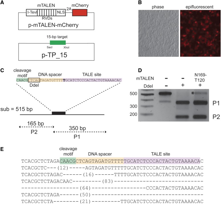
Tev-mTALEN activity in HEK293T cells on an episomal target. (A) Schematic of the vectors used for co-transfection experiments. For the expression vector, the Tev-mTALEN gene is separated from the mCherry translation reporter by a T2A peptide. (B) Example of Tev-mTALEN expression vector transfection efficiency and expression in HEK293T cells, with bright field image on the left and the epifluorescent image (1-sec exposure) of the same field of view on the right. (C) Schematic of the TP15 target, with a DdeI restriction site and sizes of DdeI digestion products indicated. (D) Agarose gel of a representative assay in which the target region was amplified by PCR from total DNA isolated 48 hr after transfection, and products were digested with DdeI (+) or incubated in buffer without DdeI (−); product resistant to cleavage by DdeI, which would result from mutagenic repair after cleavage by the mTALEN, is labeled with an asterisk (*). (E) Examples of mutations in the target resulting from co-transfection with N169-T120 or D184-V152 Tev-mTALENs. The DdeI-resistant product from (C) was cloned and several clones were sequenced. Dashes indicate the length of deletions observed in individual clones relative to the wild-type sequence.

