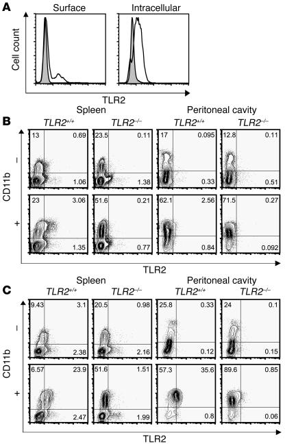
Figure 5.
TLR2 expression ex vivo immediately after primary cell isolation. Flow cytometry of splenocytes and peritoneal washout cells from wild-type (TLR2+/+) and TLR2–/– mice ex vivo immediately after isolation (n = 5, cells pooled for each sample). (A) CD11b+ splenocytes from mice challenged with LPS for 24 hours were analyzed for surface and intracellular TLR2 expression by staining with T2.5 (bold line, TLR2+/+; filled area, TLR2–/–). (B and C) For analysis of TLR2 regulation upon infection, mice were either left uninfected (–) or infected with B. subtilis and sacrificed after 24 hours (+). Upon staining of CD11b, cells were stained with T2.5 (TLR2) either without permeabilization (B) or after permeabilization (C). Numbers in quadrants represent the percentage of single- or double-stained cells with respect to the total number of viable cells analyzed.

