Figure 4.
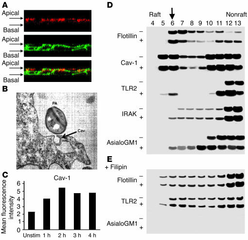
Lipid rafts are involved in clustering of receptors and signaling. (A) Confocal z-section images demonstrate caveolin-1 labeled with Alexa Fluor 594 (red) and GM1, identified with cholera toxin β-subunit (CTB) conjugated to Alexa Fluor 488 (green), on the apical surfaces of polarized 16HBE cells permeabilized after stimulation with Pam3Cys-Ser-Lys4. (B) CF nasal polyp cells were infected with P. aeruginosa PAO1 (PA) and grown on semipermeable supports, and transmission electron micrograph were obtained after 3 hours of bacterial exposure. Arrow (Cav) indicates the flask-shaped electron-dense structures typical of caveolae (magnification, ∞30,000). (C) Flow cytometry was used to detect superficial caveolin-1 on polarized 16HBE cells after exposure to S. aureus. Unstim, unstimulated. (D) Aliquots of Triton-insoluble lysates of 16HBE cells obtained before (–) and after (+) stimulation with P. aeruginosa PAO1 were fractionated on discontinuous sucrose gradients (4–40%) and were immunoblotted with anti-flotillin, anti-caveolin, anti-TLR-2, anti-IRAK-1, or anti-asialoGM1. Downward arrow indicates raft fraction containing all the components after stimulation. (E) Aliquots of sucrose gradient fractions from cells treated with filipin prior to stimulation with P. aeruginosa PAO1 were immunoblotted with anti-flotillin, anti-TLR2, and anti-asialoGM1.

