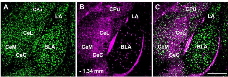
Figure 1.
Distribution of Green Fluorescent Protein (GFP) containing neurons in the amygdala of a GAD67-GFP knock-in mouse. NeuN-ir (A) labels the entire neuronal population. Large numbers of GFP-ir neurons (B) are present throughout the central amygdala, while much smaller numbers are seen in the basolateral amygdala, and lateral amygdala. Panel C contains a merged image. Abbreviations: BLA – basolateral amygdala, CeC – centrocentral, CeL – centrolateral amygdala, CeM – centromedial amygdala, CPu – caudate putamen, LA – lateral amygdala. – 1.46 mm to bregma. Scale bar = 200 μm.

