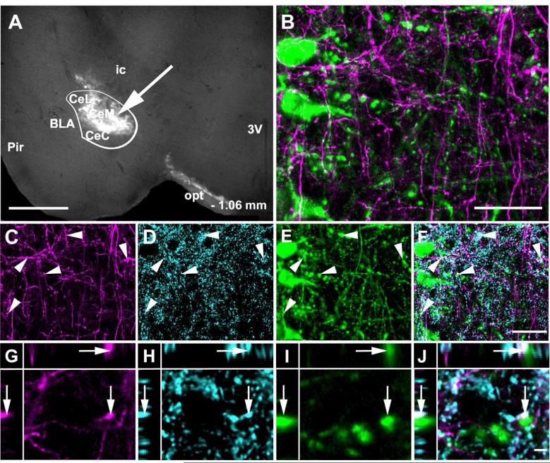
Figure 13.
Channel rhodopsin 2 (ChR2).Venus adeno-associated virus injected into the CAmy labels fibers in the LC. Panel A shows an injection site (arrow) that is concentrated in the centromedial amygdala. The white line indicates the CAmy. Panel B shows ChR2-Venus labeled fibers in the Peri-LC zone that are close to TH-ir positive neurons and fibers. ChR2-Venus fibers (C) colocalize with GAD65/67-ir (D) and are dispersed among TH-ir dendrites (E). Areas of overlap are indicated in the merged image (F, arrowheads). Possible colocalization of all three markers (ChR2-Venus; GAD65/67-ir; TH-ir) in a single optical section is shown in panels G (ChR2-Venus), H (GAD65/67-ir), I (TH-ir) and in J as a merged image. The image is presented as a XYZ image (larger panel X-Y, with Y-Z left and X-Z top). The arrow points to a puncta with possible colocalization between ChR2-Venus, GAD65/67ir and TH-ir. Abbreviations: 3V – third ventricle, BLA – basolateral amygdala, CeL – centrolateral amygdala, CeM – centromedial amygdala, CeC – centrocentral amygdala, ic – internal capsule, opt – optic tract, Pir – piriform cortex. – 1.06 mm indicates distance to bregma. Scale bar = 500 μm for A, 20 μm from B to F and scale bar = 5 μm for G to K.

