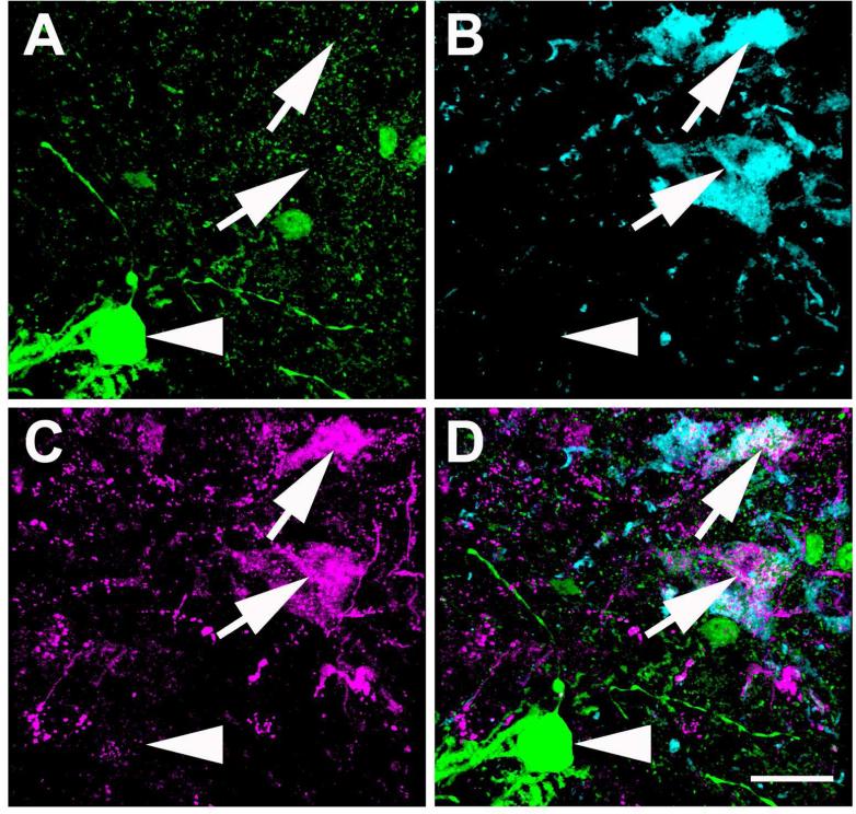
Figure 15.
High magnification image of LC TH-ir neurons labeled with HSV1-H129-LStdT after injection in the CAmy of a VGAT-iCre/GFP mouse. Panel A shows GFP expression, panel B shows TH-ir positive neurons, panel C shows HSV1-H129-LStdT labeled cells and panel D is a merged image. Arrows point to neurons that coexpress TH-ir and tdTomato from HSV1-H129-LStdT. The arrowhead points to a neighboring GABAergic neuron without a viral expression. Scale bar = 20 μm.

