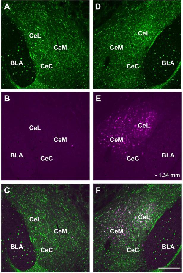
Figure 4.
The CAmy to LC projection is strictly ipsilateral. Panels A, B, and C show the right side and panels D, E and F the left side of a single coronal section from an animal injected with FG into the left LC. Only cells in the left CAmy contain significant FG signal. Panels A and D show GAD67-GFP-ir neurons, panels B and E show FG-ir cells. Panels C and F contain merged images. Abbreviations: BLA – basolateral amygdala, CeC – centrocentral amygdala, CeL – centrolateral amygdala, CeM – centromedial amygdala. – 1.34 mm indicates distance from bregma. Scale bar = 200 μm.

