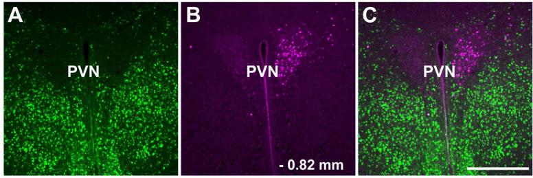
Figure 8.
Distribution of FG containing cells in the hypothalamic paraventricular nucleus after injection into LC. Panel A shows GAD67-GFP-ir neurons in the paraventricular nucleus and anterior hypothalamus, panel B shows FG labeled paraventricular neurons and panel C is a merged image. The retrogradely labeled cells in the paraventricular nucleus do not contain GAD67-GFP-ir. Scale bar = 200 μm. Abbreviations: PVN – paraventricular hypothalamic nucleus.

