Figure 3.
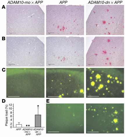
Detection and quantitation of amyloid plaques in brains from 17- to 19-month-old (A_D) APP[V717I] transgenic mice, double-transgenic ADAM10-mo ∞ APP[V717I] mice, and ADAM10-dn ∞ APP[V717I] mice. Immunohistochemical detection of amyloid plaques in the neocortex in paraffin-embedded sections with either antibody 6F/3D (A) or antibody 4G8 (B). Note that in ADAM10-mo ∞ APP[V717I] mice, no additional plaques were detected by antibody 4G8. Arrows point to blood vessels. Scale bars: 200 ∝m. (C and E) Thioflavine S_stained β structures in the subiculum of either 17- to 19-month-old (C) or 12-month-old mice (E); scale bars: 200 ∝m. (D) Quantitation of amyloid load in subiculum in thioflavine S_stained sections obtained from 17- to 19-month-old animals. Surface thioflavine S staining is expressed as a percentage of the total subiculum surface. Statistical analysis was performed for each genotype with the following number of animals: APP[V717I], n = 5; ADAM10-mo ∞ APP[V717I], n = 13; ADAM10-dn ∞ APP[V717I], n = 6. *P < 0.05; **P < 0.01.

