
Figure 2.
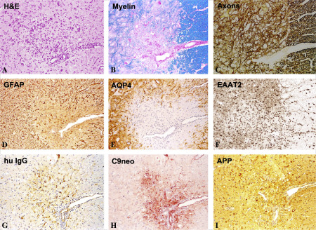
Pathology of experimental neuromyelitis optica (NMO) in the rat. Activation of myelin basic protein (MBP) reactive T‐cells by immunization with MBP/complete Freund's adjuvant (CFA) and intraperitoneal injection of human aquaporin 4 antibody containing NMO‐immunoglobulin G (IgG), beginning at the first signs of clinical symptoms and repeated 24 h later. The animals were sacrificed 48 h after the first NMO‐IgG application. As in NMO lesions in humans, the lesions are infiltrated by lymphocytes and macrophages (A), there is some loss of myelin (B), but no loss of axons in the early stage (C); astrocytes, reactive for glial fibrillary acidic proteins (GFAP) are partially lost in the lesions (D) and there is even more widespread loss of aquaporin 4 (E); the excitatory amino acid transporter 2 (EAAT2) is also lost in areas of aquaporin 4 loss (F); within active lesions there is deposition of human immunoglobulin (G) and activated complement (C9neo antigen; H); immunocytochemistry for amyloid precursor protein shows axonal spheroids in the lesions, indicating ongoing axonal injury (I); × 60. APP = amyolid precursor protein; H&E = hematoxylin and eosin.