
Fig. 1.
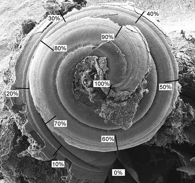
Method of measuring the length of the cochlear duct to make comparisons in corresponding positions. In order to compare hair cell bundles in corresponding positions along cochlear ducts between wildtype and mutant mice or between different mutant mice, the images were taken under a standard magnification (× 80) to show the whole organ of Corti. The cochlear duct was divided into ten parts of 10% from base to apex. A higher magnification (× 15 k) was used for analysis of IHCs and the outermost row of OHCs at every 10% interval along the length of the cochlear duct. Scale bar, 300 μm.