Fig. 10.
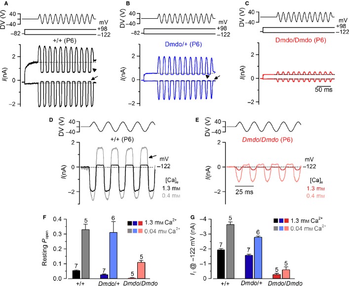
Mechanotransducer currents in diminuendo cochlear outer hair cells. (A–C) Saturating transducer currents recorded from (A) a control (B), a heterozygous and (C) a homozygous mutant P6 apical-coil diminuendo OHC by applying sinusoidal force stimuli of 50 Hz to the hair bundles at −122 mV and + 98 mV. The driver voltage (DV) signal of ± 40 V to the fluid jet is shown above the traces (negative deflections of the DV are inhibitory). The holding potential was −82 mV. Extracellular Ca2+ concentration was 1.3 mm. The arrows and arrowheads indicate the closure of the transducer currents (i.e. resting current) elicited during inhibitory bundle displacements at hyperpolarized and depolarized membrane potentials, respectively. Note that the resting current increases with membrane depolarization. Dashed lines indicate the holding current, which is the current at the holding membrane potential. (D and E) Comparison of transducer currents recorded from (D) a control and (E) a homozygous mutant P6 diminuendo OHC in the presence of 1.3 mm Ca2+ (black/red) and endolymphatic-like Ca2+ concentration (0.04 mm; grey/pink lines) at −122 mV. (F and G) Resting current (F) and (G) peak transducer current at a membrane potential of −122 mV recorded in OHCs from the three genotypes in the presence of 1.3 mm (black/blue/red) and 0.04 mm (grey/pale blue/pink) extracellular Ca2+.

