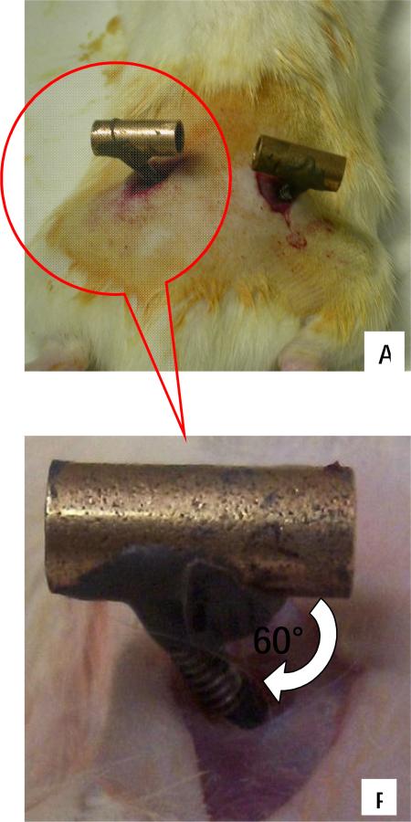
Figure 3. Surgical Images showing pelvic muscles and orthotic placement made through small incisions (Dorsal Aspect).
A) Image showing each half of the implanted pelvic orthosis from dorsal aspect. The aluminum rod is then used to connect and clamp the two halves together. B) Insert from ‘A’ showing close up of inserted screw rod of orthosis through small opening in gluteus

