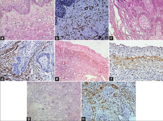
Figure 2.

(a) Odontogenic keratocyst (OKC) showing cystic lining epithelium and connective tissue capsule (H&E stain, ×400). (b) Photomicrograph showing á-SMA positive myofibroblasts in the cyst wall of OKC (IHC stain, ×400). (c) Follicular ameloblastoma showing odontogenic epithelial islands in connective tissue stroma (H&E stain, ×400). (d) Photomicrograph showing á-SMA positive myofibroblasts around odontogenic epithelial islands in follicular ameloblastoma (IHC stain, ×400). (e) uminal variant of unicystic ameloblastoma (H&E stain, ×200). (f) Photomicrograph showing á-SMA positive myofibroblasts in unicystic ameloblastoma (IHC stain, ×400). (g) Malignant epithelial islands in well-differentiated oral squamous cell carcinoma (H&E stain, ×400). (h) Photomicrograph showing á-SMA positive myofibroblasts around tumor islands in oral squamous cell carcinoma (IHC stain, ×400)
