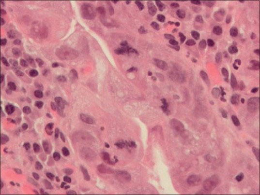
Figure 4.

Photomicrograph of gastric mucosa with high grade dysplasia (H&E stain, ×400) Three clearly identifiable mitoses are seen. The one at the 12:00 o’clock position is tripolar, that is, atypical. The mitoses at 3:00 and 6:00 o’clock are normal. Acute (neutrophils) and chronic (plasma cells and lymphocytes) inflammatory cells are in the lamina propria immediately underneath of the dysplastic epithelium. Atypical mitoses are a characteristic of precancerous lesions, that is, dysplasia and malignancy, that is, cancer. (Coutesy: En.wikipedia)
