Figure 8.

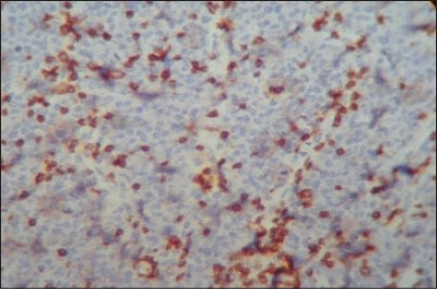
Photomicrograph showing small cell T-lymphocytes positive for CD3 with intense nuclear brown staining for cell membrane surface (IHC stain, ×400)

Photomicrograph showing small cell T-lymphocytes positive for CD3 with intense nuclear brown staining for cell membrane surface (IHC stain, ×400)