Abstract
Raine syndrome is a rare genetic disorder with characteristic features of exophthalmos, choanal atresia or stenosis, osteosclerosis and cerebral calcifications. Most of babies with this disorder die immediately after birth. We report a baby who was 7 weeks old at the time of presentation.
Keywords: Lethal, osteosclerosis, Raine syndrome
Introduction
Raine syndrome, also called as lethal osteosclerotic bone dysplasia, first described by Raine and Winter in 1989 in a term female baby, which died soon after birth. This has high mortality and most babies are still born or die during the neonatal period. This is very rare genetic disorder and very few cases have been reported all over. To the best of our knowledge, this second reported case from India.
Case Report
A 1-month-20-day-old baby, born to second degree consanguineous parents, was brought with complaints of inability to suck at the breast and failure to thrive since birth. There was a history of episodes of cyanosis while feeding, which used to subside by crying. Baby was fed with cow's milk and expressed breast milk. Mother was booked a case with no significant antenatal history. The baby cried soon after birth. This was fifth baby born to parents. The first baby and fourth baby died soon after birth and they had features suggestive of Raine syndrome. Third child is 4-year-old now and he is healthy.
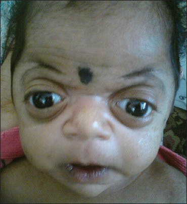
Physical examination revealed stable vitals and there was no cyanosis. Baby was weighing 2.75 kg, length was 48 cm and head circumference was 33 cm. The anterior fontanel was bulged; it was measuring 6 cm × 4 cm and posterior fontanel 5 cm × 4 cm. There was exophthalmos, small nose with depressed nasal bridge, gum hyperplasia, high-arched narrow palate, long philtrum and dysplastic ears [Figure 1]. Baby was also having short fingers and toes. The infant feeding tube (sizes 5 and 6) could not be passed through the nose indicating associated choanal atresia/stenosis. Fibreoptic posterior rhinoscopy examination revealed the presence of choanal atresia. Baby's lungs were clear cardiovascular system, central nervous system and abdominal examinations were normal.
Figure 1.

Raine syndrome baby showing exophthalmos, small nose with depressed nasal bridge, long philtrum and dysplastic ears
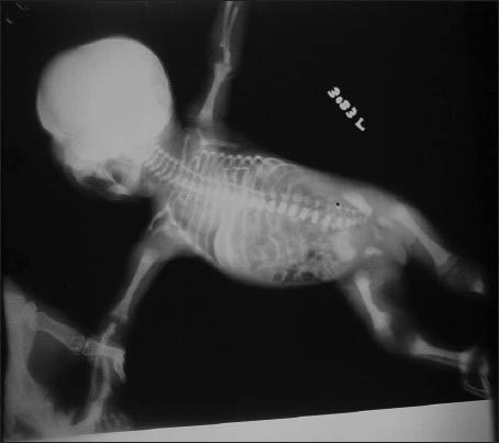
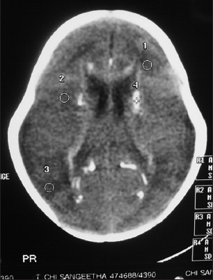
Baby's routine blood investigations were normal. Infantogram of the baby showed generalized osteosclerosis [Figure 2]. Computed tomography scan of the brain showed diffuse hyperdense areas of calcification in periventricular region, corpus callosum and basal ganglia on both sides [Figure 3]. Serology for congenital cytomegalovirus infection was negative. The molecular studies could not be carried out due to the financial constraints. Diagnosis of Raine syndrome was made based on typical clinical presentation, radiographic picture and brain tomography findings.
Figure 2.

Infantogram of baby showing generalized osteosclerosis of bones
Figure 3.

Computed tomography scan of the brain showing calcifications in periventricular white matter, corpus callosum and basal ganglia
Discussion
Raine syndrome, also known as lethal osteosclerotic bone dysplasia, is a rare autosomal recessive disorder characterized by exophthalmos, microcephaly, depressed nasal bridge, bilateral choanal atresia/stenosis, gum hyperplasia and osteosclerosis.[1] The osteosclerosis is usually generalized, but occasionally may be focal involving only few bones.[2] Palate usually shows cleft, but may be higharched and narrow. The babies affected with this disorder show intracranial calcifications.[3] Calcifications are seen in parietal and occipital periventricular white matter and corpus callosum, but never occur in cortex, temporal lobes, internal capsule, cerebellum and brain stem.[4]
Major association between sclerosing dysplasia and intracranial calcification is seen in osteopetrosis associated with renal tubular acidosis and carbonic anhydrase II deficiency.[5] However, in this disorder, the calcifications are usually seen after 2 years of age exclusively in basal ganglia and cortex and characteristic clinical features of Raine syndrome are lacking.[3,6]
It is inherited as autosomal recessive; mutations in FAM20C gene on chromosome 7 are identified in babies with this disorder.[7]
Initial reports showed that this disorder is lethal and most of babies with this disorder died in early months of their life. However, mutations in FAM20C gene were also noticed in older children with phenotypic features of Raine syndrome with osteosclerosis, indicating that lethality is not essential for diagnosis.[8]
Footnotes
Source of Support: Nil
Conflict of Interest: None declared.
References
- 1.Raine J, Winter RM, Davey A, Tucker SM. Unknown syndrome: Microcephaly, hypoplastic nose, exophthalmos, gum hyperplasia, cleft palate, low set ears, and osteosclerosis. J Med Genet. 1989;26:786–8. doi: 10.1136/jmg.26.12.786. [DOI] [PMC free article] [PubMed] [Google Scholar]
- 2.Al-Gazali LI, Jehier K, Nazih B, Abtin F, Haas D, Sadagahatian R. Further delineation of Raine syndrome. Clin Dysmorphol. 2003;12:89–93. doi: 10.1097/00019605-200304000-00003. [DOI] [PubMed] [Google Scholar]
- 3.Al Mane KA, Coates RK, McDonald P. Intracranial calcification in Raine syndrome. Pediatr Radiol. 1996;26:55–8. doi: 10.1007/BF01403707. [DOI] [PubMed] [Google Scholar]
- 4.Rickert CH, Rieder H, Rehder H, Hülskamp G, Hörnig-Franz I, Louwen F, et al. Neuropathology of Raine syndrome. Acta Neuropathol. 2002;103:281–7. doi: 10.1007/s00401-001-0469-5. [DOI] [PubMed] [Google Scholar]
- 5.Ohlsson A, Cumming WA, Paul A, Sly WS. Carbonic anhydrase II deficiency syndrome: Recessive osteopetrosis with renal tubular acidosis and cerebral calcification. Pediatrics. 1986;77:371–81. [PubMed] [Google Scholar]
- 6.Nampoothiri S, Anikster Y. Carbonic anhydrase II deficiency a novel mutation. Indian Pediatr. 2009;46:532–4. [PubMed] [Google Scholar]
- 7.Simpson MA, Hsu R, Keir LS, Hao J, Sivapalan G, Ernst LM, et al. Mutations in FAM20C are associated with lethal osteosclerotic bone dysplasia (Raine syndrome), highlighting a crucial molecule in bone development. Am J Hum Genet. 2007;81:906–12. doi: 10.1086/522240. [DOI] [PMC free article] [PubMed] [Google Scholar]
- 8.Simpson MA, Scheuerle A, Hurst J, Patton MA, Stewart H, Crosby AH. Mutations in FAM20C also identified in non-lethal osteosclerotic bone dysplasia. Clin Genet. 2009;75:271–6. doi: 10.1111/j.1399-0004.2008.01118.x. [DOI] [PubMed] [Google Scholar]


