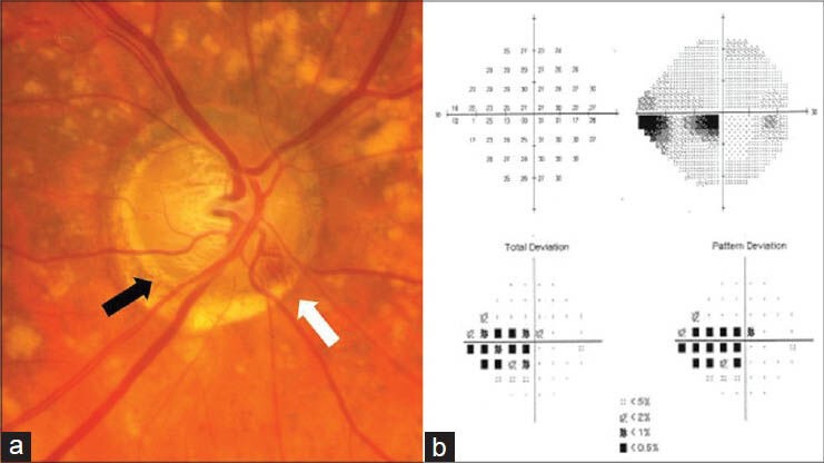
Figure 1.

(a) Right optic nerve with an inferotemporal acquired pit of the optic nerve indicated by the black arrow. A disc hemorrhage is also seen in the inferonasal region (white arrow). (b) Standard automatedperimetry shows a corresponding superior paracentral arcuate defect with an inferior paracentral arcuate secondary to superior rim narrowing
