
Figure 1.
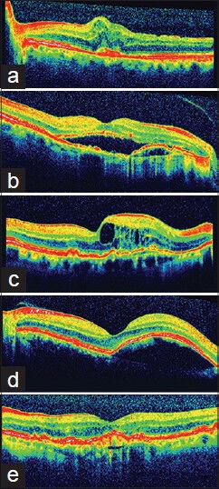
Spectral domain optical coherence tomography features of choroidal neovascularization, by which the authors qualified patients for anti-VEGF treatment. (a) Increased retinal thickness without subretinal and/or intraretinal fluid (b) Subretinal fluid (c) Intraretinal fluid (d) Pigment epithelium detachment (e) Fibro vascular pigment epithelium detachment