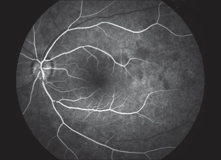
Figure 3.

Fundus fluorescein angiography of the LE. Fluorescein angiography showed a patchy background choroidal flourescence and a relative delay in arterio-venous filling in the superior temporal retinal vessels compared with the inferior temporal ones in the early phase. No early hyperfluorescence of the disc or vessels was observed
