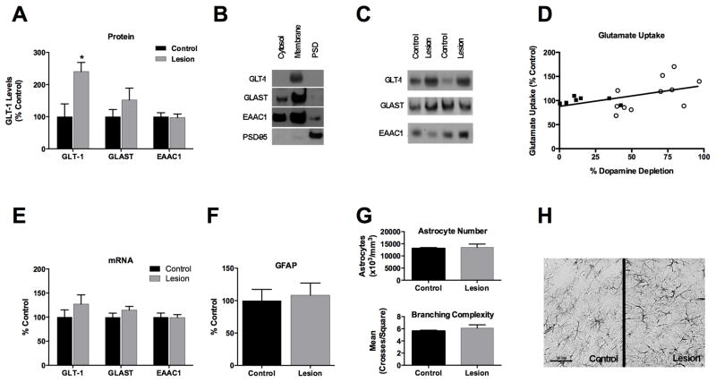
Figure 1.
A). Dopamine loss in the PFC increases GLT-1 protein levels in the membrane- enriched fraction; there is no significant change in GLAST or EAAC1. B). Glutamate transporters are most abundant in the membrane-enriched fraction. C). Representative immunoblot showing the glutamate transporters in control and dopamine-depleted (lesion) animals. D). The magnitude of the decrease in PFC dopamine is positively correlated to the degree to which glutamate uptake is increased. E). There is no change in mRNA levels of GLT-1 or other glutamate transporters. F). No change in level of the astrocytic protein GFAP is seen in immnoblot studies, and G) stereological evaluation indicates no increase in the number of GFAP-positive astrocytes (top) or astrocytic processes (bottom) in dopamine- denervated animals. H). Representative immunohistochemical staining for GFAP; scale bar = 50μm.
*p = .011

