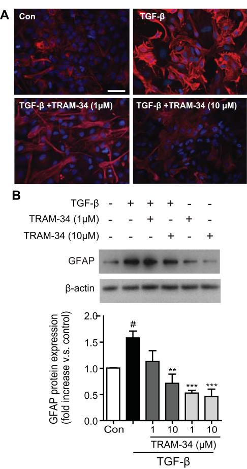
Fig. 2.
Blockade of KCa3.1 channel attenuates TGF-β-induced GFAP protein expression. (A) Representative immunocytochemistry showing GFAP expression in cultured astrocytes treated with TGF-β (10 ng/ml) for 5 days in the presence of TRAM-34 (1, 10μM). Scale bar = 50 μm. Con: control. (B) Representative western blot showing GFAP expression in cultured astrocytes treated with TGF-β (10 ng/ml) for 5 days in the presence of TRAM-34 (1, 10μM). Quantification of western for GFAP immunoreactivity (n = 3). Data are presented as means ± SEM. #p < 0.05 versus control, **p < 0.01, ***p < 0.001 versus TGF-β alone.

