


Figure 7.
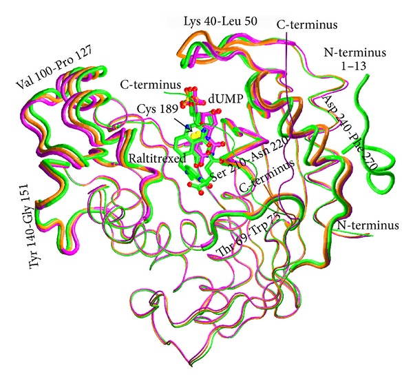
Superimposition of the main chains of the three structures: ligand-free mTS (orange, PDB ID: 3IHI), mTS-dUMP complex (violet, PDB ID: 4E5O), and mTS-dUMP-Raltitrexed complex (green, PDB ID: 4EB4). Ligands, dUMP and Raltitrexed, and catalytic Cys189 are shown as sticks. N, O, and S atoms are colored blue, red, and yellow, respectively. C atoms are colored the same as the corresponding tubes representing the main chains of respective complexes. The most differing main chain fragments are shown as thicker tubes. The 13-residue N-terminal fragment (1–13) in the tertiary complex, having no equivalents in the ligand-free enzyme and the binary complex, is also shown.