
Figure 6. Lithium attenuates the anti-angiogenic and anti-Wnt effects of kallistatin.
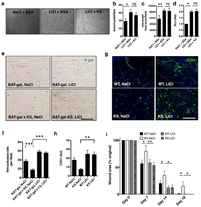
(a) Tube formation assay with HDMVECs; (b) total branch points, n=3; (c) total tube length, n=3; (d) TCF/β-catenin transcriptional activity in HDMVECs; (e) X-gal staining showing Wnt-activation in day 7 wounds of Wnt reporter mice in response to NaCl or LiCl topical treatments. (f) Quantification, Wnt-reporter X-gal activity; (g) CD31 signaling in day 7 wound beds after topical treatments; (h) quantification, CD31+ cells, day 7 wound beds (AU=arbitrary fluorescence units); (i) Overall skin wound healing rate expressed by wound area; N=7-10 age-matched male mice; n=5 or >5. Mean±S.E.M *p<0.05; **<0.01; ***p<0.001; ANOVA and Tukey’s post-hoc significance analysis performed. Scale bars (e, g): 50 μm.