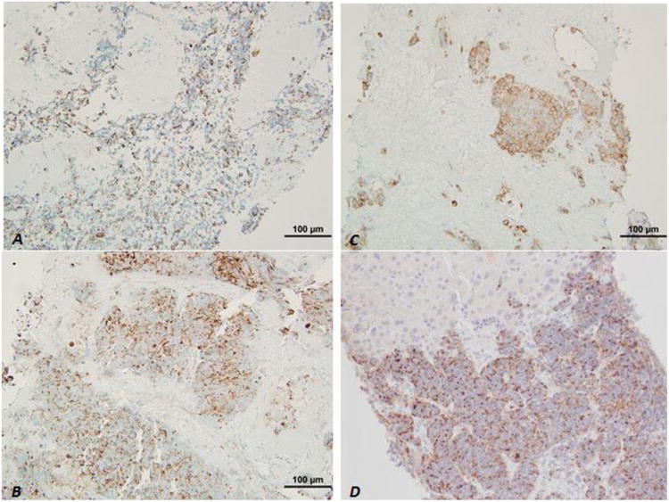
Figure 4.

Immunohistochemistry of liver core biopsies. Neoplastic cells in both the nodular and diffuse patterns were positive for AE1/AE3 (A), and residual hepatocytes were positive for CK Cam5.2 (B); and, neoplastic cells were also highlighted by synaptophysin (C) and chromogranin (D).
