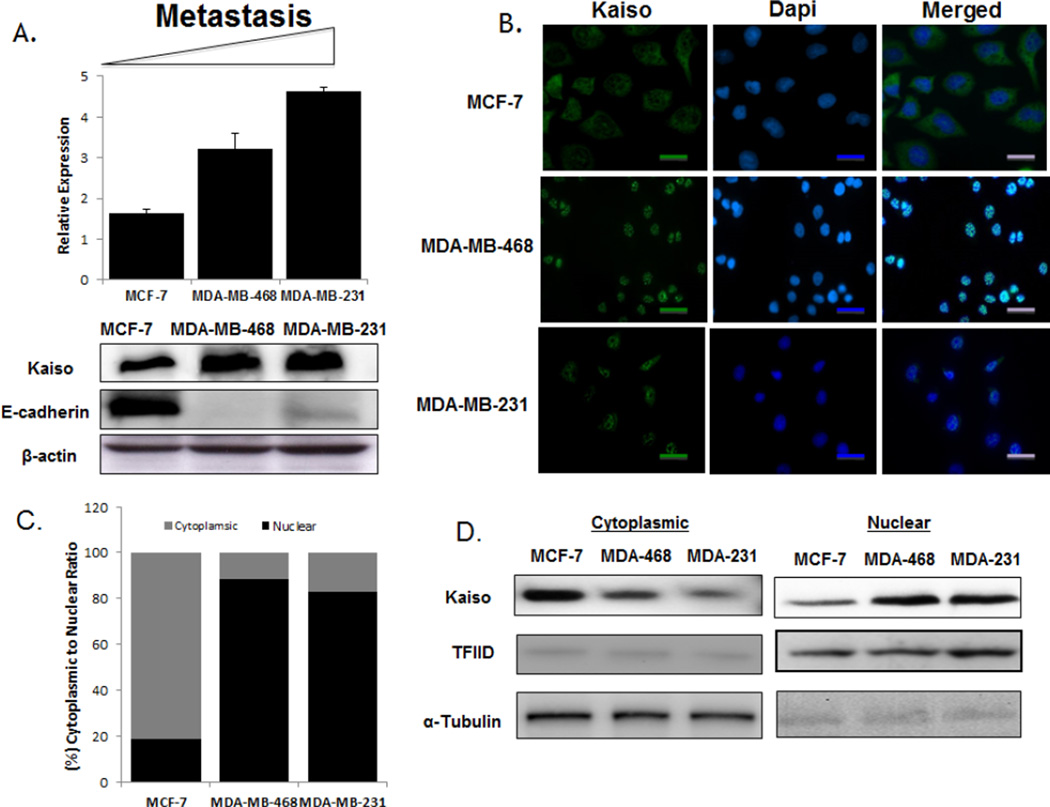
Fig. 4.
Kaiso expression and localization in breast cancer cell lines. (A) Kaiso mRNA expression levels, measured by real-time PCR, revealed that increasing expression correlated with aggressiveness for MCF-7, MDA-MB-468, and MDA-MB-231 cells. Immunoblot analyses of cells treated with the anti-Kaiso 6F8 antibody showed increasingly higher expression of Kaiso protein in MCF-7, MDA-MB-468, and MDA-MB-231 cells, correlating with less to more aggressive cell lines. (B) Kaiso protein expression and localization among breast cancer cells, determined by immunofluorescence, showed that MCF-7 cells displayed high cytoplasmic expression relative to the metastatic cell lines, MDA-MB-468 and MDA-MB-231, which have a higher nuclear expression. Anti-mouse Alexa 488 (green) was utilized as secondary antibody and DAPI as a nuclear counter-stain (blue). (C) Bar-graph quantification of Kaiso fluorescent intensity in the individual cytoplasmic and nuclear compartments. (D) Cytosolic and nuclear fractions were isolated by sequential extraction. Anti-α-tubulin was used as a loading control for the cytoplasmic fraction and TFIID as a loading control for the nuclear fraction.

