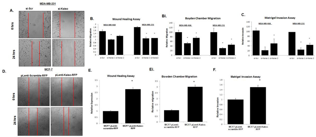
Fig. 5.
Down-regulation of Kaiso expression results in decreases of migration and invasion. (A) Cells treated with siRNA Kaiso 1, siRNA Kaiso 2, or scrambled siRNA were wounded, and cell migration was analyzed after 24 hr. Photos were taken at 100× magnification. MDA-MB-231 images serve as representative images of both MDA-MB-468 and MDA-MB-231 cells. Red vertical bars indicate the starting area migration on Day 0. (B) Quantitative analysis of relative migration also showed that MDA-MB-231 and MDA-MB-468 cells treated with siRNA Kaiso 1 or 2 had delays in migration relative to untreated cells or cells treated with scrambled siRNA (si-Scr). (Bi) Migration rates of MDA-MB-231 and MDA-MB-468 cells treated with siRNA Kaiso 1 or 2 were measured in Boyden cambers and compared to cells treated with si-Scr controls. (C) MDA-MB-468 and MDA-MB-231 cells treated with siRNA Kaiso 1 or 2 showed a decrease in the number of cells invading through a layer of Matrigel relative to control cells and cells treated with si-Scr. (D) MCF-7 cells transfected with pLenti-scramble-RFP plasmid or pLenti-Kaiso-RFP were wounded, and cell migration was measured at 24 hr. Photos were taken at 100× magnification. Red vertical bars indicate the starting area migration on Day 0. (E) Quantitative analysis of relative migration shows that MCF-7 cells treated with pLenti-Kaiso-RFP had an increased capacity to migrate relative to cells treated with the pLenti-scramble-RFP plasmid. (Ei) Migration rates of MCF-7 cells transfected with pLenti-scramble-RFP plasmid or pLenti-Kaiso-RFP were measured at 24 hr in Boyden chambers. (F) MCF-7 cells transfected with pLenti-Kaiso-RFP showed an increase in the number of cells invading through a layer of Matrigel relative to cells treated with the pLenti-scramble-RFP plasmid. All data presented are the means of three independent experiments, performed in triplicate ± s.e. *P<0.05.

