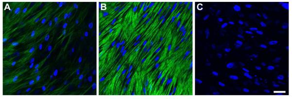
Figure 4.

Indirect immunofluorescent confocal images of collagen type III on full-thickness constructs at 4 weeks. A) Control, B) T1: TGF-β1 treated, and C) Rescue: TGF-β1 for 2 weeks then switched to TGF-β3 for 2 weeks. Type III collagen was upregulated upon T1 stimulation, when compared to Control; however, it was diminished upon Rescue treatment. Blue = TOPRO3 nuclear counterstain, Bar = 50 microns.
