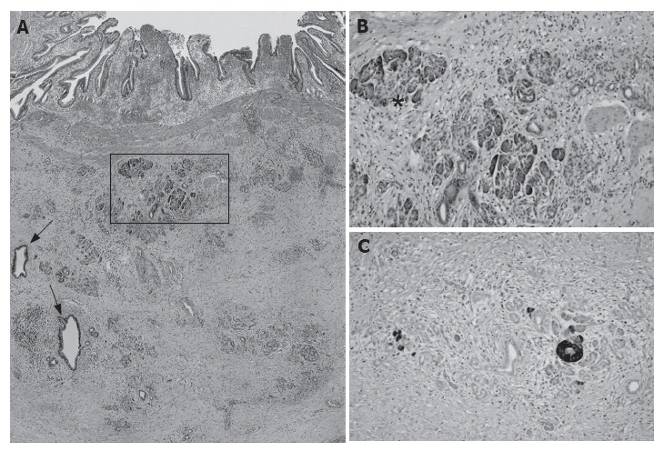
Figure 1.

Histological sections of HP showing dilated ducts (arrows, H&E x 100) and acini (A), acinar and islet cells (asterisk, H&E x 250) (B), and chromogranin A expressing islet cells (Mayer’s haematoxylin x 250) (C) in submucosa of gallbladder neck.
