
Figure 5.
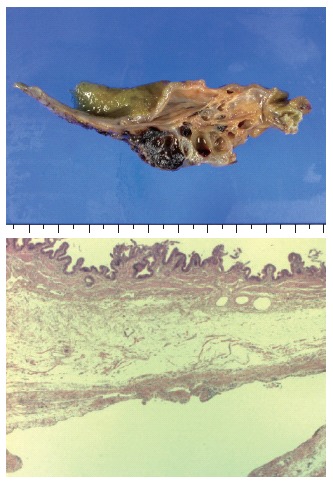
Gross appearance of resected specimen showing a well-marginated multi cystic lesion filled with yellowish fluid, and microphotograph from the cholecystectomy specimen showing a lymphatic space lined with flat endothelium (hematoxylin and eosin staining, x12.5).