
Figure 3.
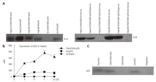
A: E1A and E1B expression identified by Western blot demonstrating that all HCC cells infected with CNHK500-p53 or WtAd5 were positive for E1A expression, however, normal BJ cells were negative for E1A expression when they were infected with CNHK500-p53, and positive only when they were infected with WtAd5, while E1B of CNHK500-p53 was only expressed under hypoxia condition in HepG2 and Hep3B, and expressed both under normal and hypoxia condition with WtAd5; B: ELISA assay showing that p53 protein secreted from a HepG2, infected with CNHK500-p53, was significantly higher than that infected with nonreplicative adenovirus Ad-p53 and Ad-Blank in vitro (P < 0.05); C: Western blot showing enhanced p53 expression in HepG2 infected with CNHK500-p53.