
Figure 2.
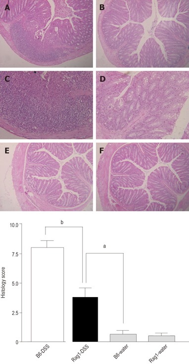
Hematoxylin and eosin (HE) staining of colons obtained from B6 and Rag-1 KO mice treated with or without DSS. A, C: B6 mice treated with DSS (X100 and 160); B, D: Rag-1 KO mice treated with DSS (X100 and 160); E: B6 mice treated with water (X100); F: Rag-1 KO mice treated with water (X100); G: Quantitative histopathologic assessment of DSS-induced colitis activity showing a significant suppression in Rag-1 KO mice compared to the B6 control mice. Samples were collected from B6 control and Rag-1 KO mice treated with 1.5% DSS (open and solid bar, respectively), or water (stripped bars). Data are expressed as mean ± SE and represent > 10 mice per group (aP<0.05; bP<0.01).