
Figure 4.
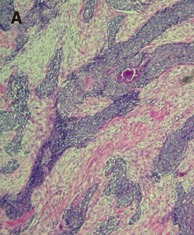
A: Photomicrography depicts nests or clusters of small tumor cells outlined by characteristic desmoplastic stroma bands (hematoxylin and eosin staining, 100×). B: Photomicrography demonstrates the monomorphic, small round tumor cell population with a small spherical or spheroid nucleus, rich in chromatin (hematoxylin and eosin staining, 400×). C: The immunohistochemical staining of tumor cells is positive for desmin.