
Figure 1.
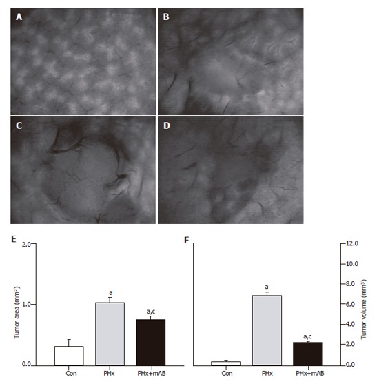
Intravital fluorescence microscopy at d 7 after tumor cell implantation. A: Displays the normal liver microvasculature; B: shows the microvasculature of the tumor of a non-resected control animal (Con); C: displays a 7-day tumor and its microvasculature of an animal which underwent hepatectomy (PHx); D: shows the tumor microvasculature after additional anti-MIP-2 treatment (PHx+mAB). Quantitative analysis of tumor area (E) and tumor volume (F) showed that hepatectomy (PHx) markedly accelerated tumor growth when compared with controls (Con), and that additional anti-MIP-2 treatment (PHx+mAB) was capable of significantly reducing this liver resection-induced tumor growth. Mean ± SE; aP < 0.05 vs Con; cP < 0.05 vs PHx. Magnifications (A-D) ×16.