
Figure 3.
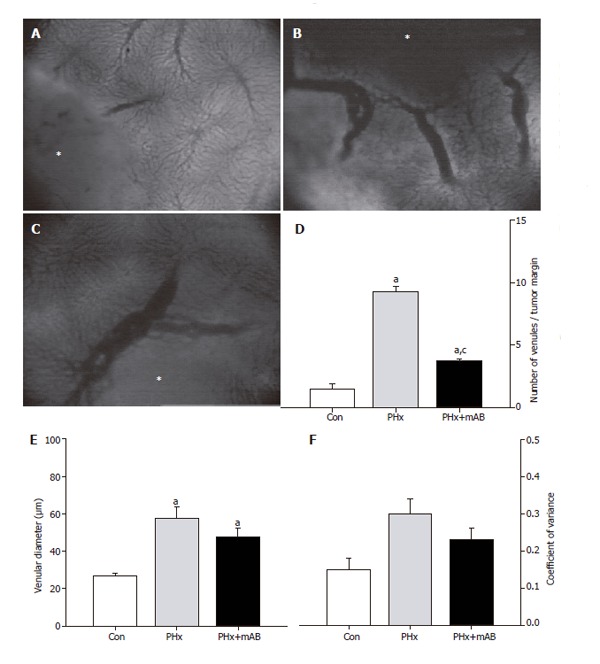
Analysis of the number of venules within the margin of tumors (A-C, asterisk) of control mice (A, Con), after hepatectomy (B, PHx) and after hepatectomy and additional anti-MIP-2 treatment (C, PHx+mAB). Quantitative analysis showed that hepatectomy significantly increases the number of venules (D), and causes a marked dilation of the draining venules within the margin of the tumors (E). Additional anti-MIP-2 treatment was capable of significantly reducing the liver resection-induced increase of the number of tumor venules (D). The heterogeneity of venular diameters, as given by the coefficient of variance, was not affected in either of the treatment groups (F). Mean ± SE; aP < 0.05 vs Con; cP < 0.05 vs PHx. Magnifications (A-C) ×40.