
Figure 4.
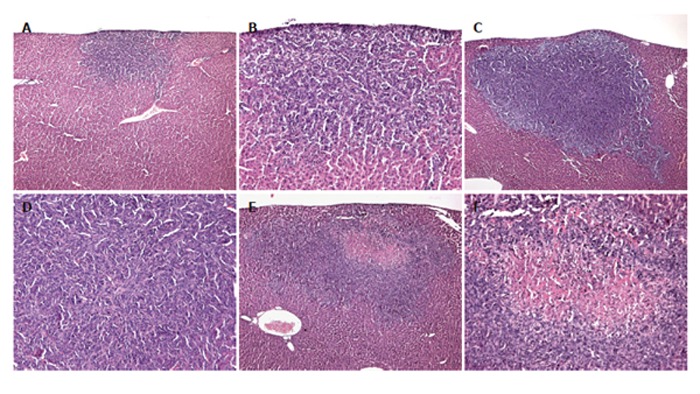
Hematoxylin–eosin-staining of tumors of a control mouse (A and B), a mouse which underwent hepatectomy (PHx) (C and D), and a mouse which additionally received anti-MIP-2 treatment (E and F). Sections revealed solid growth of the colorectal CT26.WT hepatic metastasis in all the animals. Tumors were round in nature, however, showed aggressive growth characteristics. Anti-MIP-2 treatment provoked an area of necrosis within the tumor center (E and F). Magnifications (A, C and E) ×18, (B, D and F) ×88.