
Figure 6.
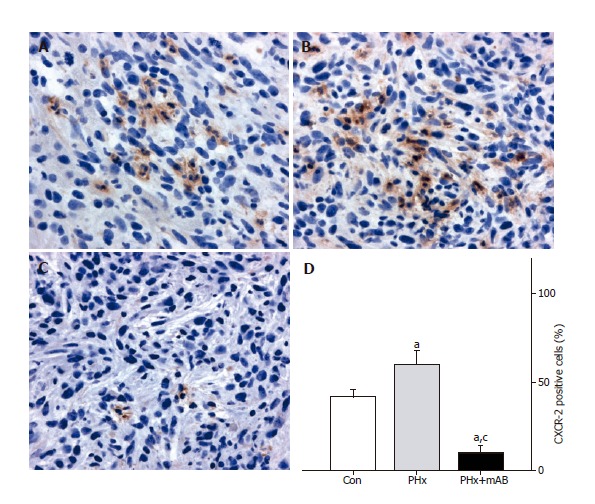
CXCR-2 immunohistochemistry and quantitative analysis of the number of receptor-positive cells (given in percent of all cells) revealed that within tumors of control animals (Con) ~40% of the cells express CXCR-2 (A and D). Hepatectomy (PHx) significantly increased CXCR-2 expression to ~60% (B and D). Of interest, additional blockade of MIP-2 (PHx+mAB) significantly reduced tumor cell CXCR-2 expression and inhibits significantly the liver resection-induced increase of CXCR-2 expression (C and D). Mean ± SE; aP < 0.05 vs Con; cP < 0.05 vs PHx. Magnifications (A-C) ×175.