
Figure 8.
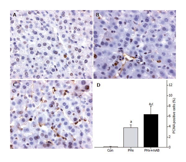
PCNA immunohistochemistry of normal liver tissue of control mice (A, Con), after hepatectomy (B, PHx), and after hepatectomy and additional anti-MIP-2 treatment (C, PHx+mAB). Quantitative analysis revealed that hepatectomy increases the number of PCNA-positive stained cells when compared to controls (D). Additional neutralization of MIP-2 further enhanced the number of PCNA-positive cells (D). Mean ± SE; aP < 0.05 vs Con; cP < 0.05 vs PHx. Magnifications (A-C) ×175.