
Figure 2.
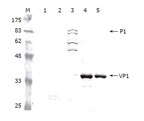
Western blot analysis of cell lysates infected by different viruses. The Sf-9 cells were infected by the viruses at a total MOI of 10 and harvested at 3 dpi. The proteins were separated by SDS-PAGE, electrotransferred to a nitrocellulose membrane and probed using anti-VP1 MAb as the primary antibody. Lane 1: mock infection; Lane 2: wild-type baculovirus AcMNPV; Lane 3: Bac-P1; Lane 4: Bac-P1-3CD; Lane 5: Bac-P1 and Bac-3CD.