
Figure 7. Brite adipocytes are Myf5/Pax3-lineage positive or negative depending upon the depot.
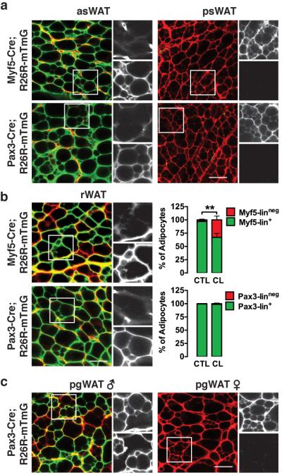
(a) Representative images of whole-mount preparations of asWAT and psWAT of 6-week old male myf5-cre;R26R-mTmG (top) and pax3-cre;R26R-mTmG (bottom) mice that were treated with CL316,243 for one week.
(b) Representative images of whole-mount preparations of rWAT from male myf5-cre;R26R-mTmG (top) and pax3-cre;R26R-mTmG (bottom) mice treated with CL316,243 for one week starting at 5 weeks of age. (Right) Quantification of mTFP+ and mGFP+ mature adipocytes in the rWAT of control (CTL) and treated (CL) mice is shown (n=4–5 mice for myf5-cre; n=4–8 mice for pax3-cre. mean+S.E.M; **, p<0.01; t-test).
(c) Representative images of whole-mount preparations of male and female pgWAT of 6-week old pax3-cre;R26R-mTmG mice that were treated with CL316,243 for one week. Scale bar= 50 μm.