FIGURE 4.
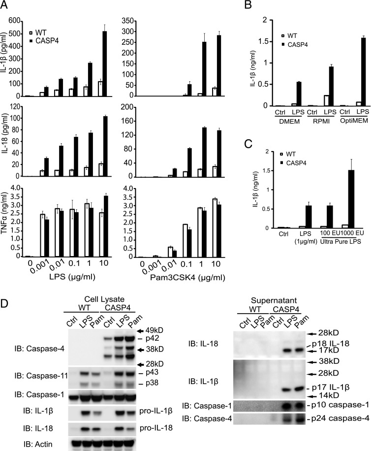
Caspase-4 mediates IL-1β and IL-18 secretion after stimulation with TLR ligands. (A) BMDMs from WT or caspase-4 transgenic (CASP4) mice were stimulated with the indicated concentration of LPS or Pam3CSK4 for 24 h, and the levels of IL-1β, IL-18, and TNF-α in supernatants were determined by ELISA. Error bars represent SEM. (B) WT or CASP4 BMDMs were treated with LPS (1 μg/ml) in the indicated medium for 24 h. IL-1β was quantitated by specific ELISA assay. (C) BMDMs from the indicated genotypes of mice were stimulated with LPS or ultrapure LPS for 24 h. Levels of IL-1β in supernatants were determined by ELISA. (D) Immunoblot (IB) analyses of lysates (left panel) and supernatants (right panel) from WT and CASP4 BMDMs treated for 24 h with control medium (Ctrl) or 1 μg/ml of LPS or Pam3CSK4 (Pam). Data representative of three independent experiments are shown.

