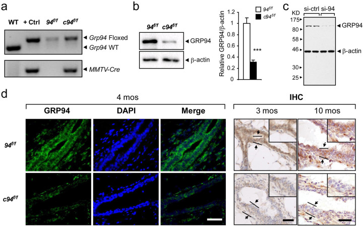
Figure 1. Generation of MMTV-Cre mediated GRP94 knockout mouse model.
(a) Representative PCR genotyping results from tails of 94f/f and c94f/f mice. The alleles are indicated. Cropped gels were shown and the full-length gels were indicated in Supplementary Fig. 2a. (b) Representative Western blot detection of GRP94 levels in mammary epithelial cells isolated from 94f/f and c94f/f glands at 2.5 months, with β-actin serving as loading control. Quantitation of GRP94 level after normalization to the β-actin level is shown on right (n = 2 per genotype). Data are presented as mean ± S.E. p < 0.001. Cropped blots were shown and the full-length blots were indicated in Supplementary Fig. 3a. (c) Western blot analysis of HBL100-HER2 cells transfected with either si-control (si-ctrl) or si-Grp94 (si-94) for detection of GRP94, with β-actin as loading control. (d) Immunofluorescent (IF) and immunohistochemical (IHC) staining of GRP94 in mammary glands from 94f/f and c94f/f mice at the indicated ages. PMSG was injected two days before euthanasia of 4 and 10 month old mice to synchronize estrous cycle. Green or brown color depicts GRP94 staining. Blue color depicts DAPI staining. Scale bars show 50 μm and are applicable to all sections. Negative controls for staining were shown in Supplementary Fig. 4a, b.

