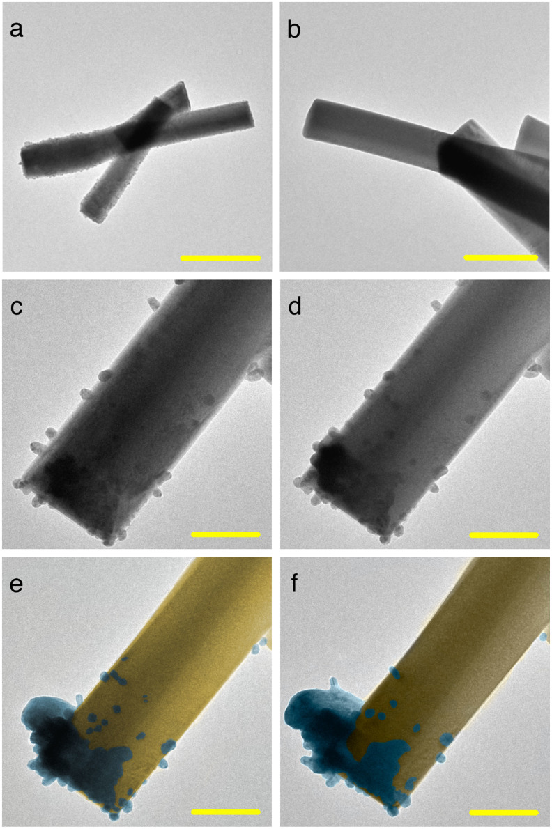
Figure 1. TEM images of the formation of Ag filaments from the a-Ag2WO4 bulk.
(a) and (b) TEM images obtained at different magnifications indicate a smooth and clear surface. (c)–(f) Thick Ag filaments grow at the edge of the sample, whereas other Ag nanoparticles are absorbed by the matrix. (Scale bar = 500 nm in a, 200 nm in b and, 100 nm in (c–f).

