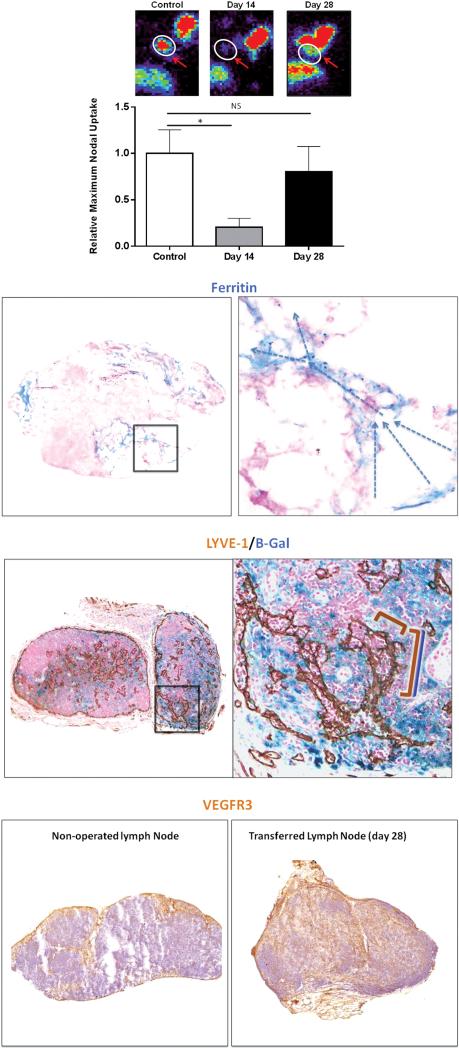
Figure 2. Lymph node transplantation restores lymphatic transport capacity and function by promoting infiltration of lymphatics from the recipient mouse.
(A) Representative heat maps (top) and quantification of relative Tc99 uptake in axillary lymph nodes after injection in the distal upper extremity of control mice (axillary incision without lymphadenectomy), and experimental mice 14 and 28 days after lymph node transplantation. Note recovery of Tc99 uptake by day 28.
(B) Representative low (left panel; 2.5×) and high (right panel; 40×) powered images of ferritin staining in lymph nodes harvested 28 days after transplantation (arrows denote direction of lymphatic flow from lymphatic sinuses toward the cortex).
(C) Representative low (left panel; 2.5×) and high powered (right panel; 40× zoom of boxed region in left panel) images of transplanted lymph nodes double stained with the lymphatic specific marker LYVE-1 (brown stain) and B-gal (blue stain). Note connection of double stained vessels (overlapping blue/brown brackets) with single stained brown vessel (brown bracket) suggesting connection of recipient and donor lymphatics within the lymph node.
(D) Representative low power (2.5×) images of control (sham-operated; left panel) and transplanted lymph node (right panel) stained for VEGF-R3 (brown stain) demonstrating massive infiltration of VEGF-R3 positive cells in transplanted node.

