Figure 1.

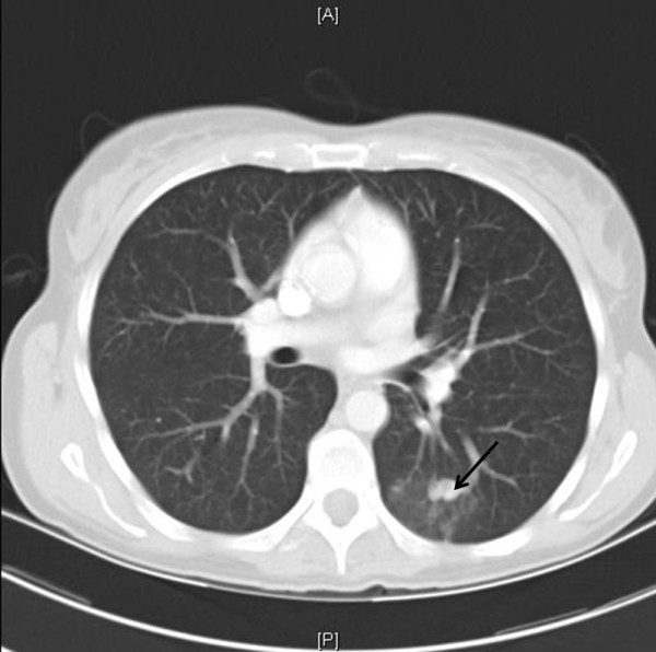
Axial computed tomography image at level of aortic root demonstrates small left lower lobe pulmonary arteriovenous malformation (arrow) with surrounding ground glass opacity consistent with recent haemorrhage.

Axial computed tomography image at level of aortic root demonstrates small left lower lobe pulmonary arteriovenous malformation (arrow) with surrounding ground glass opacity consistent with recent haemorrhage.