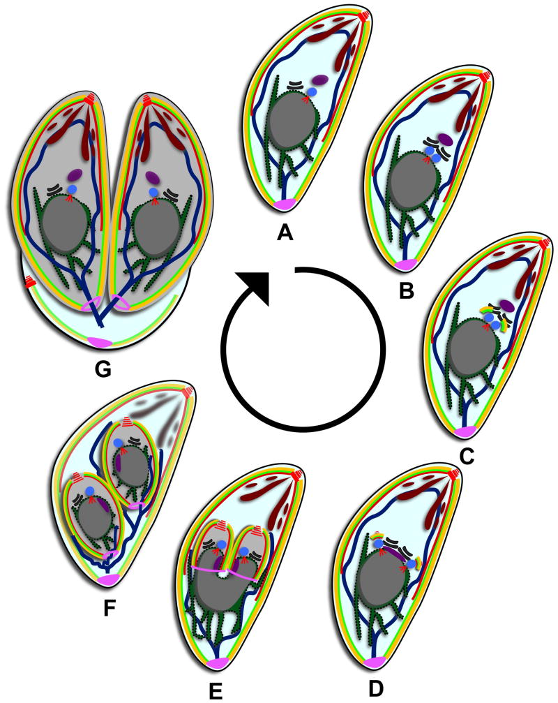
Figure 1.
Toxoplasma divides by internal daughter budding. (A) Mature parasite in G1. Red are MT (conoid, subpellicular, and spindle pole), yellow are the alveoli, bright green is the IMC meshwork, brown are secretory vesicles, dark blue line is the mitochondrion, purple is the apicoplast, blue is the centrosome, black is the Golgi apparatus, dark green is the ER, grey is the nucleus, and pink is the posterior cup or basal complex. (B) Mitosis is initiated at 1.2N with the duplication of the Golgi apparatus followed by the duplication of the centrosomes. (C) Budding is initiated with the appearance of early components of the cytoskeleton. (D) The spindle pole duplicates and the apicoplast moves below the centrosomes, elongating as the centrosomes separate. (E) The organelles begin to partition as the daughter buds elongate. The components of the basal complex accumulate at the leading edge of the bud. (F) The daughter buds contract and all the organelles are partitioned except for the mitochondrion. The secretory vesicles and cytoskeleton of the mother begin to degrade. (G) Daughter buds emerge and the plasma membrane from the mother is incorporated onto the daughters. The mother falls away as a residual body.

