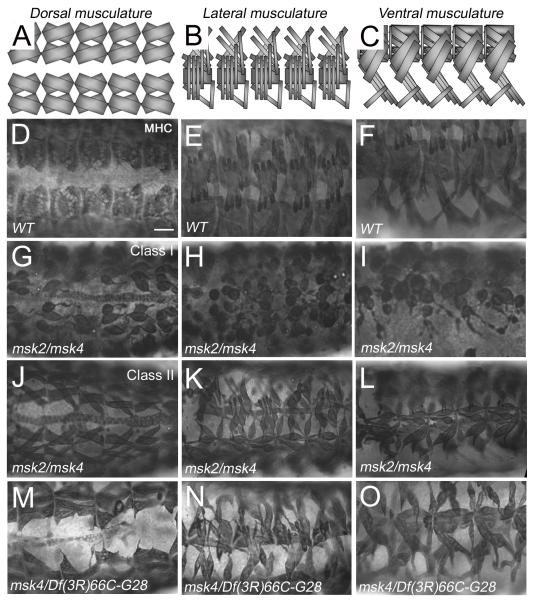
Figure 1. Msk mutant embryos exhibit defects in somatic muscle attachment.
(A-C) Schematic illustration of the WT muscle pattern in the dorsal (A), lateral (B) and ventral (C) musculature. (D-O) The dorsal (D, G, J, M), lateral (E, H, K, N), and ventral (F, I, L, O) musculature in stage 17 embryos visualized immunohistochemically with MHC antibody. (D-F) Wild-type embryos exhibit a stereotypical repeating pattern of muscles in each hemisegment. (G-I) Embryos trans-heterozygous for the msk2 and msk4 alleles exhibit severe muscle attachment defects (Class I) that affect all muscle subsets (12.1%; n=33). (J-L) The trans-heterozygous combination msk2/msk4 shows variable penetrance as 45.5% of the embryos show moderate defects in muscle attachment (Class II). (M-O) msk4 over a deficiency that removes the msk locus also exhibits muscle attachment defects with variable penetrance. In all embryos, anterior is to the left. Scale bar: 20 μm.

