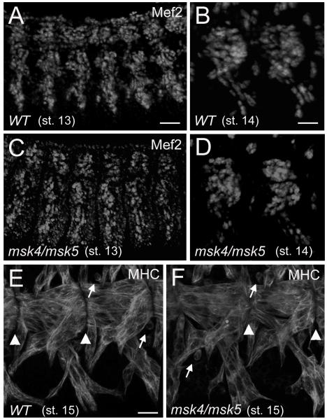
Figure 2. Muscle cell fate and myotube migration are not affected in msk mutant embryos.
(A-D) Immunofluorescent stainings of stage 13 (A, C) and stage 14 (B, D) embryos. (A-D) Similar numbers of Mef2-expressing myoblasts are observed in WT (A, B) and msk4/msk5 homozyogous embryos (C, D) in both the lateral (A, C) and ventral longitudinal muscles (B, D). (E, F) Confocal micrographs of the ventral musculature in stage 15 embryos stained with a monoclonal antibody to MHC. (E) By stage 15 in WT embryos, the ventral longitudinal muscles have migrated to the site of muscle-tendon attachment. (F) The ability of the ventral muscles to find their proper attachment sites is not affected in msk mutants. Arrowheads denote muscle-tendon attachment sites and arrows point to unfused myoblasts still present in stage 15 embryos. In all embryos, anterior is to the left and dorsal is up. Scale bars: 50 μm in A, C; 20 μm in B, D; 10 μm in E, F.

