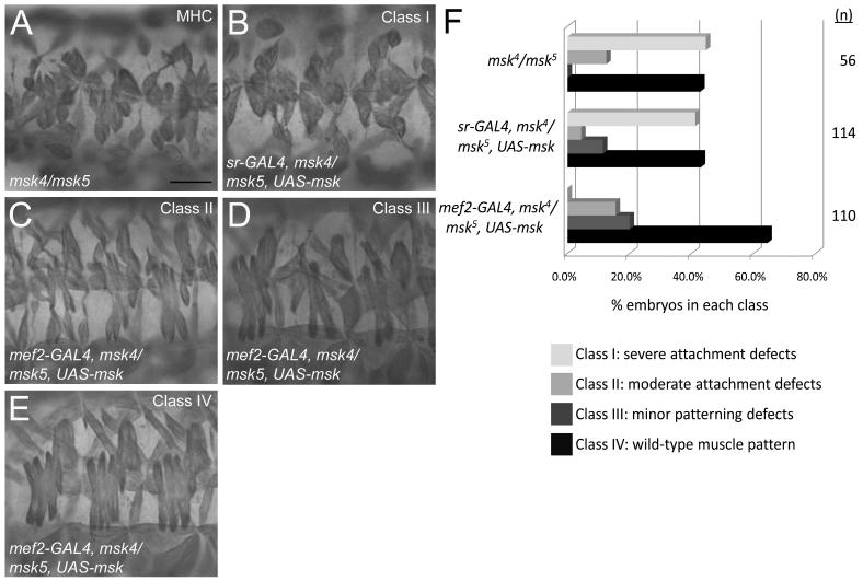
Figure 3. Msk is required in muscle cells for proper muscle-tendon attachment.
(A-E) MHC staining of 3 hemisegments of the lateral musculature in stage 17 embryos. (A) A representative embryo of the genotype msk4/msk5 shows severe defects in muscle attachment where muscles have pulled away from attachment sites and are found in a “ball.” (B-E) Expression of Msk in either the tendon cells (B) or the muscle cells (C-E) in msk4/msk5 embryos using the GAL4/UAS system. (B) Expression of UAS-msk in the tendon cells using sr-GAL4 does not rescue the muscle attachment defects (Class I). (C-E) Expression of UAS-msk in the muscle under control of the mef2 promoter results in minor attachment defects (C; Class II), minor patterning defects (D; Class III), or rescue to a WT muscle pattern (E; Class IV). Scale bar: 20 μm (F) A bar graph quantitating the ability of mef2-GAL to either partially (Class II; 15.5%) or fully (Class IV; 64.5%) rescue the muscle attachment defects in msk4/msk5 embryos (n=110). In contrast, embryos with sr-GAL4 are not rescued by expression of Msk in the tendon cells and exhibit phenotypes quantitatively similar to that of msk4/msk5 mutants only. Scale bar: 20 μm.

