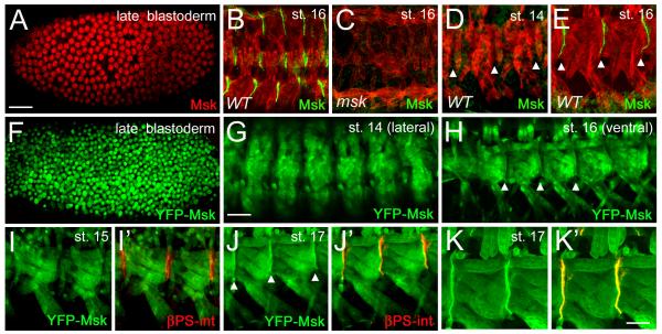
Figure 4. Msk protein localizes to muscle-tendon attachment sites in late stage embryos.
(A-E) Msk protein expression and localization in embryogenesis visualized using anti-Msk antisera. (A) Msk protein is nuclear in late blastoderm embryos. (B-E) Msk protein is in green and muscle is labeled in red using anti-Tropomyosin (TM). (B) Msk becomes enriched at the dorsal, lateral and ventral muscle-tendon attachment sites in stage 16 embryos. (C) Msk attachment site staining is absent in msk4/msk5 mutant embryos. (D, E) Msk protein is not observed at the ventral muscle-attachment sites in stage 14 embryos (D), but is observed after stage 15 (E). (F-K’) Msk protein expression visualized using the fusion protein Msk-YFP (green). Expression of MTD-GAL4::Msk-YFP in late blastoderm embryos (F) and mef2-GAL4::Msk-YFP in stage 16 embryos (G-K’). The Msk-YFP protein expression and localization (F) mimics the nuclear Msk protein expression (A). YFP-Msk protein is detected in both the nucleus and cytoplasm in stage 14 (G) and stage 16 (H) embryos, but becomes enriched at the ends of muscles in the latter stage (arrowhead in H). (I’, J’, K’) Double labeling with βPS-integrin (red) shows the location of the muscle attachment site in the ventral musculature. YFP-Msk expression occurs throughout the muscle, but is not enriched near βPS-integrin in stage 15 embryos (Fig. 4I, I’). In stage 17 embryos, YFP-Msk protein is enriched at the sites of muscle attachment and colocalizes with βPS-int (Fig. 4J’, K’; arrowheads). Scale bars: 50 μm in A-D; 20 μm in E-H’; 10 μm in I-I’.

